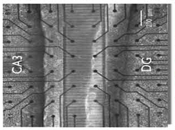
The neurnal activity between the dentate gyrus (DG) and CA3 subregions of the brain are of particular interest for understanding memory-impaired diseases like Alzheimer's. To study the activity dynamics, we are engineering a MEMs based platform, allowing repeatable in vitro studies with 3D cultured cells.
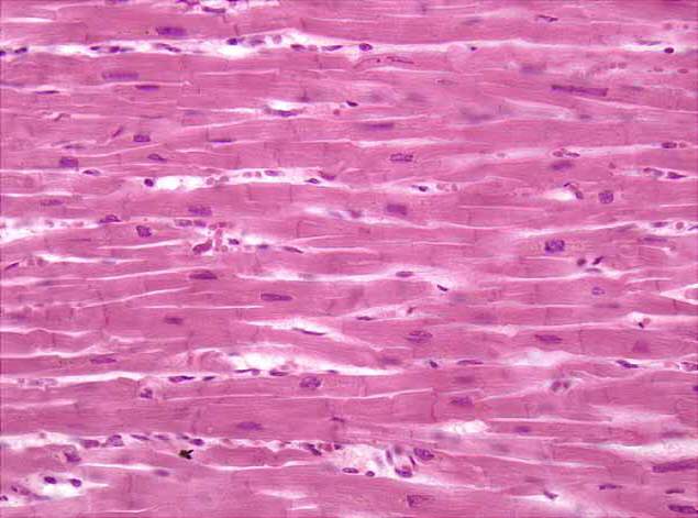
Prior experimental results showed that micron-size ridges and grooves correlate with an increased orientation of cultured cardiomyocytes. Various patterns with micron-sized precision were created using PDMS molded from a master formed from soft-lithography. Primary cells from neonatal rats were grown in these fabricated PDMS platforms. Quantification of cell alignment was classified by: percented of oriented cells and ratio of major/minor axis of the fitted ellipses over cells. Results showed neonatal rate ventricilar myoctyes responded more strongly to envirionmental structures than HL-1 cells. (description adapted from 2018 conference poster by Lee, C.; Agnew, W.J.; and Tang, W.C.)

The goal of this project is to develop a hardware-software codesigned platform to analyze the physiological parameters of a preterm infant in Neonatal Intensive Care Unit (NICU). One of the key factors affecting preterm infant care and eventual discharge is oral feeding readiness. This platform, Pedi~Sync, collects physiological data before, during, and after oral feeding and analyzes the breathing pattern with a custom developed algorithm to generate a health score, which is then further integrated with other physiological data to establish care regimen and time to discharge.
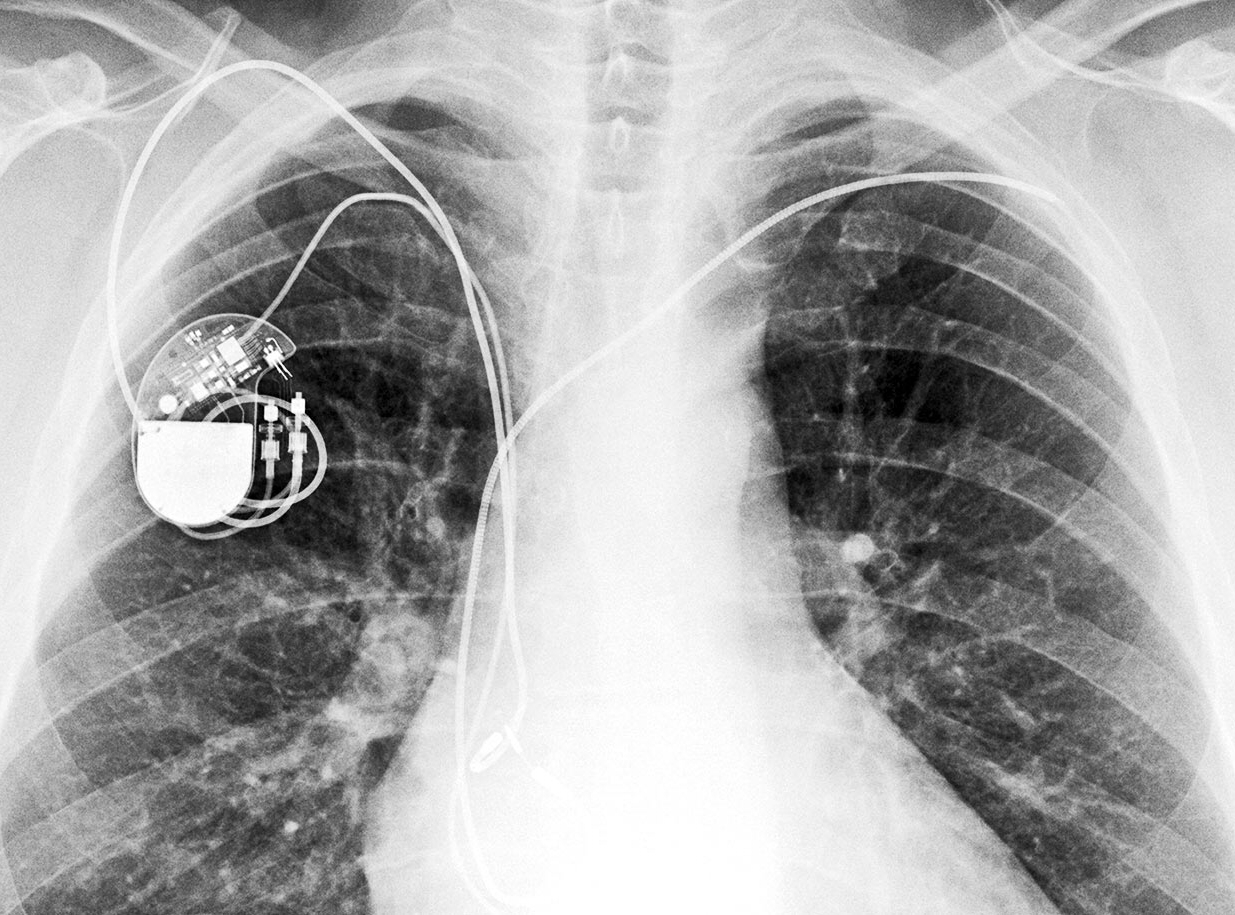
The aim of this project is to develop an energy-harvesting device that can be integrated with a typical leadless pacemaker without increasing the volume to extend the device’s lifespan by at least twofold to 15 years or longer. MEMS technology would be a prime candidate to implement the miniature energy harvester. The operating mechanism is to convert the periodic contractile energy of heart muscles or pulsatile blood flow into electricity, which can then be used to trickle charge an onboard rechargeable battery.
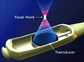
Alzheimer’s disease (AD) is a growing epidemic with approximately 44 million people diagnosed worldwide. Common symptoms include memory loss, agitation, and difficulty with planning or problem solving. During the time between mild and severe cognitive impairment, there is a significant accumulation of the hallmark biomarker, amyloid beta (Aβ). Extracellular Aβ plaques are associated with neuronal damage, brain atrophy and volume reduction leading to the life-threatening symptoms. Currently, there is no cure for the disease and treatment options like pharmaceutical and cognitive stimulation are costly, ineffective, and inconsistent. The purpose of this project is to engineer and test the feasibility transcranial focused ultrasound (tFUS) device that exhibits the potential for treating AD.
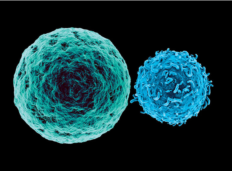
Tumor cells can go unrecognized by our immune system, leading to malignant growths and the development of cancer. As a result, on this platform, we try to reactivate immune cells, specifically T-cells, to recognize and attack tumor cells. In addition, the micro-platform, will permit observation of the interaction between cancer cells and immune cells, i.e. cell migration, cytosis and so on. This device may also provide immediate feedback on the effectiveness of cancer immunotherapy. In the future work, this microdevice can be used for precision medicine and finding the optimal immune treatment for every patient.
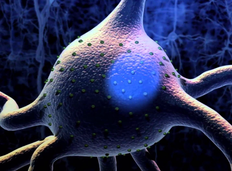
Neuronal network activity allows an individual to perceive, think, and react to environment. When these activities are disrupted, our ability to respond to outside stimuli may be compromised. One of the common method of studying these activity is observing neuron in vitro on microelectrode array(MEA). However, the spatiotemporal resolution can be compromised in this setting due to crosstalk of electrical stimuli. To address this issue, we are using optogenetics, where we use certain wavelength of light to stimulate specific neuron, to observe neuron network activity without the risk of crosstalk.
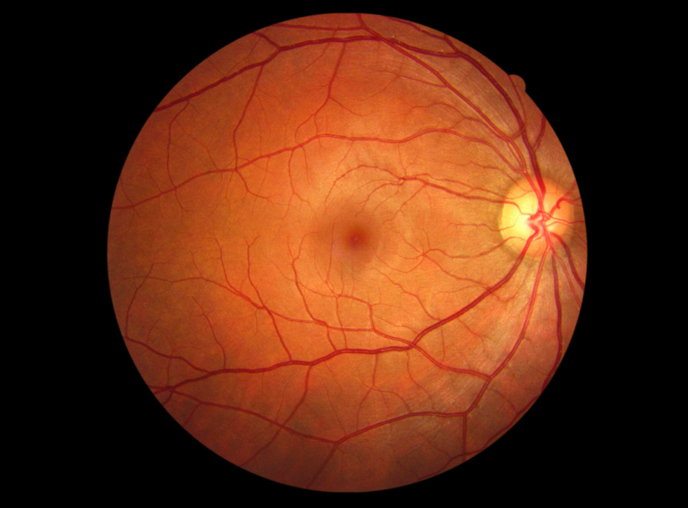
Under specific culture conditions, human pluripotent stem cells will differentiate into a certain type of cells, which would further self-organize into biological structures that mimic natural organs. These structures, called organoids, have great potentials for transplantation therapy of different retinal diseases. The goal of this project is to design and create a diffusion only microfluidic platform to culture an already-created retinal organoid for an extended period of time, and eventually grow organoids with consistent and good quality for future transplantation. This platform would eliminate the influence of mechanical shear stress and allow fluorescence lifetime imaging microscopy to monitor the metabolic activity of organoids.